Necrostatin-1
别名: Nec-1
Necrostatin-1 (Nec-1)是一种特异性RIP1 (RIPK1)抑制剂,抑制TNF-α诱导的细胞坏死,在293T细胞中EC50为490 nM。Necrostatin-1也可抑制 IDO、细胞自噬和凋亡。
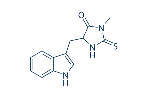
Necrostatin-1 Chemical Structure
CAS: 4311-88-0


产品质控
批次:
纯度:
99.97%
99.97
Necrostatin-1相关产品
细胞实验数据示例
| 细胞系 | 实验类型 | 给药浓度 | 孵育时间 | 活性描述 | 文献信息 |
|---|---|---|---|---|---|
| L929 | Growth Inhibition Assay | 2/5 μg/ml | 24 h | reverses the cell growth inhibition and cell death induced by TNFα alone as well as TNFα + zVAD | 23941769 |
| L929 | Function Assay | 2 μg/ml | 24 h | promots caspase-6 (p20) activity and procaspase-6 cleavage | 23941769 |
| L929 | Function Assay | 5 μg/ml | 24 h | blocks zVAD induced necroptosis and autophagy | 23941769 |
| C6 | Cell Viability Assay | 1 mmol/L | 3 h | attenuates Shikonin induced glioma cell death | 23840441 |
| U87 | Cell Viability Assay | 1 mmol/L | 3 h | attenuates Shikonin induced glioma cell death | 23840441 |
| C6 | Cytotoxicity Assay | 1 mmol/L | 3 h | blocks shikonin induced necrosis | 23840441 |
| U87 | Cytotoxicity Assay | 1 mmol/L | 3 h | blocks shikonin induced necrosis | 23840441 |
| C6 | Function Assay | 1 mmol/L | 1.5-3 h | suppresses the expression of RIP-1 caused by shikonin | 23840441 |
| U87 | Function Assay | 1 mmol/L | 1.5-3 h | suppresses the expression of RIP-1 caused by shikonin | 23840441 |
| TE671 | Cell Viability Assay | 40 μg/ml | 24 h | rescues GX15-070-induced loss of cell viability | 23744296 |
| RMS13 | Cell Viability Assay | 40 μg/ml | 24 h | rescues GX15-070-induced loss of cell viability | 23744296 |
| MEFs | Cytotoxicity Assay | 2/6/20 μM | 18 h | inhibits TNFα-induced cell death in RelA KO MEFs | 23727581 |
| MEFs | Function Assay | 20 μM | 1/2/4 h | suppresses TNFα-induced RIPK1 phosphorylation | 23727581 |
| ΔN-Karpas 299 | Cytotoxicity Assay | 20 μM | 16 h | inhibits CD30-induced cell death | 23545938 |
| MM.1S | Cytotoxicity Assay | 90 µM | 1 h | blocks BAY 11-7082 induced rapid cell swelling | 23527154 |
| KMS-12-BM | Cytotoxicity Assay | 90 µM | 1 h | blocks BAY 11-7082 induced rapid cell swelling | 23527154 |
| HT-22 | Cell Viability Assay | 10 μM | 12 h | protects against glutamate-induced cell death | 23307752 |
| HT-22 | Function Assay | 25 μM | 0–30 min | inhibits ERK Activation induced by glutamate | 23307752 |
| NIH3T3 | Function Assay | 10/50 μM | 1/3 h | ameliorates TNFα-driven complex formation | 23261677 |
| SH-EP | Apoptosis Assay | 10 μM | 72 h | inhibits IAP inhibitor- and Lexatumumab-induced apoptosis | 22890322 |
| HL60 | Apoptosis Assay | 60 μM | 12 h | enhances shikonin-induced apoptosis | 22837689 |
| HL60/Adr | Apoptosis Assay | 60 μM | 12 h | enhances shikonin-induced apoptosis | 22837689 |
| K562 | Apoptosis Assay | 60 μM | 12 h | enhances shikonin-induced apoptosis | 22837689 |
| K562/Adr | Apoptosis Assay | 60 μM | 12 h | enhances shikonin-induced apoptosis | 22837689 |
| HL60 | Function Assay | 60 μM | 12 h | augments the caspase-3 activity | 22837689 |
| HL60/Adr | Function Assay | 60 μM | 12 h | augments the caspase-3 activity | 22837689 |
| K562 | Function Assay | 60 μM | 12 h | augments the caspase-3 activity | 22837689 |
| K562/Adr | Function Assay | 60 μM | 12 h | augments the caspase-3 activity | 22837689 |
| HL60 | Function Assay | 60 μM | 12 h | increases the activity of caspases, caspase 8 and 9 | 22837689 |
| HL60/Adr | Function Assay | 60 μM | 12 h | increases the activity of caspases, caspase 8 and 9 | 22837689 |
| K562 | Function Assay | 60 μM | 12 h | increases the activity of caspases, caspase 8 and 9 | 22837689 |
| K562/Adr | Function Assay | 60 μM | 12 h | increases the activity of caspases, caspase 8 and 9 | 22837689 |
| L929sA | Apoptosis Assay | 10 μM | 1 h | inhibits the apoptotic response to TNF | 22362767 |
| L929sA | Apoptosis Assay | 10 μM | 1 h | rescues cells expressing RIPK1ΔID from TNF-induced apoptosis | 22362767 |
| L929sA | Apoptosis Assay | 10 μM | 1 h | abrogates the interaction of caspase-8 with FADD | 22362767 |
| TPC-1 | Cell Viability Assay | 100 μM | 24 h | increases cellular survival | 22136818 |
| 8505c | Cell Viability Assay | 100 μM | 24 h | increases cellular survival | 22136818 |
| SW13 | Cell Viability Assay | 100 μM | 24 h | increases cellular survival | 22136818 |
| Jurkat | Cytotoxicity Assay | 50/ 100/200 μm | 1/3 h | reduces Naegleria fowleri-induced cytotoxicity | 21535020 |
| Jurkat | Function Assay | 200 μm | 30 min | reduces Naegleria fowleri-induced reactive oxygen species (ROS) generation | 21535020 |
| HT-22 | Cytotoxicity Assay | 10 μM | 12 h | protects against cell death induced by 5 mmol/L glutamate | 17760869 |
| L929 | Function Assay | 2/5 μg/ml | 24 h | reversed the autophagy induced by TNFα alone as well as TNFα + zVAD | 23941769 |
| NRK-52E | Cell Viability Assay | 20 μM | 24 h | inhibits increased Drp1 protein expression after TNF-α Stimulation and ATP Depletion | 24351845 |
| NRK-52E | Cell Viability Assay | 20 μM | 24 h | increases cell viability after TNF-α Stimulation and ATP Depletion | 24351845 |
| NRK-52E | Cell Viability Assay | 20 μM | 24 h | protects cells from cell death caused by ischemia injury | 24351845 |
| AGS | Cell Viability Assay | 60 μm | 1 h | prevents shikonin-induced cell death | 24463199 |
| L-540 | Cell Viability Assay | 60 μm | 1 h | reduces the Givinostat/Sorafenib-induced cell death | 24561519 |
| L-540 | Function Assay | 60 μm | 1 h | prevents the mitochondrial membrane depolarization | 24561519 |
| L-540 | Function Assay | 60 μm | 1 h | prevents the generation of ROS | 24561519 |
| SK-Hep1 | Function Assay | 60 μM | 18 h | blocks β-lapachone-mediated PAR accumulation and AIF translocation to the cytosol | 24832602 |
| SK-Hep1 | Function Assay | 60 μM | 18 h | inhibits β-Lapachone-induced leakage of HMGB-1 | 24832602 |
| SK-Hep1 | Function Assay | 60 μM | 18 h | blocks β-lapachone-induced morphological change, cell death and PI uptake | 24832602 |
| Huh7 | Cell Viability Assay | 50 µM | 24/48 h | prevents cell death of rAdHCV co-infected Huh7 cells | 24973240 |
| L929 | Cell Viability Assay | 30 μM | 1 h | inhibits TNF-α-induced cleavage of Topo I | 25095742 |
| L929 | Cell Viability Assay | 30 μM | 1 h | inhibits TNF-α-induced loss of cell viability | 25095742 |
| L929-A | Function Assay | 50 μM | 12 h | inhibits the TNFα-induced loss of mitochondrial membrane permeability | 25398540 |
| L929 | Function Assay | 50 μM | 12 h | inhibits TNFα-induced Bid cleavage | 25398540 |
| L929-N | Function Assay | 50 μM | 12 h | blocks the cleavage of Caspase-3 and PARP | 25398540 |
| L929-A | Function Assay | 50 μM | 12 h | blocks the cleavage of Caspase-3 and PARP | 25398540 |
| L929-N | Cell Viability Assay | 50 μM | 24 h | blocks TNFα-induced cell death | 25398540 |
| L929-A | Cell Viability Assay | 50 μM | 24 h | blocks TNFα-induced cell death | 25398540 |
| KMS-12-PE | Cell Viability Assay | 60 μM | 5 h | inhibits SHK-induced cell death | 25530098 |
| SGC-7901 | Cell Viability Assay | 30 μM | 1 h | suppresses oxaliplatin-mediated cell death | 25767076 |
| BxPC-3 | Function Assay | 20 μM | 24 h | decreases the early necrotic cells | 26000607 |
| MiaPaCa-2 | Function Assay | 20 μM | 24 h | decreases the early necrotic cells | 26000607 |
| NCI-H28 | Cell Viability Assay | 10 μM | 24 h | prevents DAPE-induced reduction of NCI-H28 cell viability | 26004138 |
| BMDM | Function Assay | 10 μM | 30 min | protects cells from TAKI-induced LDH release | 26381601 |
| MEFs | Cell Viability Assay | 10 μM | 48 h | inhibits zVAD-promoted death of CNOT3-depleted MEFs | 26437789 |
| A549 | Cell Viability Assay | 50 μM | 24 h | inhibits MMS-induced cell death | 26472723 |
| Jurkat T | Necroptosis assay | 30 uM | 24 hrs | Inhibition of necroptosis in TNF-alpha-induced human Jurkat T cells assessed as cell viability at 30 uM after 24 hrs | 18467094 |
| L929 | Necroptosis assay | 30 uM | 24 hrs | Inhibition of necroptosis in zVAD-induced mouse L929 cells assessed as cell viability at 30 uM after 24 hrs | 18467094 |
| L929 | Necroptosis assay | 30 uM | 24 hrs | Inhibition of necroptosis in TNF-alpha-induced mouse L929 cells assessed as cell viability at 30 uM after 24 hrs | 18467094 |
| Jurkat | Cytoprotective assay | 30 uM | 1 hr | Cytoprotective activity against FasL-induced necroptosis in human Jurkat cells assessed as increase in cell viability at 30 uM incubated for 1 hr followed by FasL stimulation measured after 20 hrs by Alamar blue assay | 29541357 |
| Jurkat | Cytoprotective assay | 30 uM | 1 hr | Cytoprotective activity against CHX-induced necroptosis in human Jurkat cells assessed as increase in cell viability at 30 uM incubated for 1 hr followed by CHX stimulation by Alamar blue assay | 29541357 |
| Jurkat | Cytoprotective assay | 30 uM | 1 hr | Cytoprotective activity against Z-VAD-induced necroptosis in human Jurkat cells assessed as increase in cell viability at 30 uM incubated for 1 hr followed by Z-VAD stimulation by Alamar blue assay | 29541357 |
| Jurkat | Cytoprotective assay | 30 uM | 1 hr | Cytoprotective activity against FasL-induced necroptosis in human Jurkat cells assessed as increase in cell viability at 30 uM incubated for 1 hr followed by FasL stimulation measured after 20 hrs by phase contrast microscopy | 29541357 |
| Jurkat | Cytoprotective assay | 30 uM | 1 hr | Cytoprotective activity against CHX-induced necroptosis in human Jurkat cells assessed as increase in cell viability at 30 uM incubated for 1 hr followed by CHX stimulation by phase contrast microscopy | 29541357 |
| Jurkat | Cytoprotective assay | 30 uM | 1 hr | Cytoprotective activity against Z-VAD-induced necroptosis in human Jurkat cells assessed as increase in cell viability at 30 uM incubated for 1 hr followed by Z-VAD stimulation by phase contrast microscopy | 29541357 |
| OHC | Function Assay | 300 μM | increases the number of apoptotic OHCs without altering the levels of CC8 after noise exposure | 24874734 | |
| OHC | Function Assay | 300 μM | diminishes noise-induced AMPK activation | 24874734 | |
| OHC | Function Assay | 300 μM | results in a reduction of noise-induced RIP1 and RIP3 immunofluorescence | 24874734 | |
| OHC | Function Assay | 300 μM | decreases noise-induced swollen nuclei | 24874734 | |
| OHC | Function Assay | 300 μM | increases noise-induced condensed nuclei | 24874734 | |
| Sf9 | Function assay | 30 mins | Inhibition of recombinant human GST-fused RIPK1 (1 to 497 residues) expressed in baculovirus infected insect Sf9 cells in presence of 32P-gamma-ATP after 30 mins by autoradiogram-based Western blot method, IC50 = 0.182 μM. | 28280261 | |
| 3T3 | Cell death assay | 24 hrs | Inhibition of death receptor signaling mediated necrotic cell death in mouse 3T3 cells assessed as cell viability after 24 hrs by ATP based viability assay in presence of TNFalpha and zVAD.fmk | 16408008 | |
| 3T3 | Cell death assay | 24 hrs | Inhibition of death receptor signaling mediated necrotic cell death in mouse 3T3 cells assessed as cell viability after 24 hrs by ATP based viability assay in presence of FasL and zVAD.fmk | 16408008 | |
| MEF | Cell death assay | 16 hrs | Inhibition of death receptor signaling mediated necrotic cell death in SV40 transformed mouse MEF cells assessed as cell viability after 16 hrs by ATP based viability assay in presence of TNFalpha, CHX and zVAD.fmk | 16408008 | |
| L929 | Cell death assay | 24 hrs | Inhibition of death receptor signaling mediated necrotic cell death in mouse L929 cells assessed as cell viability after 24 hrs by ATP based viability assay in presence of TNFalpha | 16408008 | |
| U937 | Cell death assay | 48 hrs | Inhibition of death receptor signaling mediated necroptotic cell death in human U937 cells assessed as cell viability after 48 hrs by ATP based viability assay in presence of TNFalpha and zVAD-fmk | 16408008 | |
| 3T3 | Cell death assay | 24 hrs | Inhibition of death receptor signaling mediated necroptotic cell death in mouse 3T3 cells assessed as cell viability after 24 hrs by ATP based viability assay in presence of TNFalpha and zVAD-fmk | 16408008 | |
| Jurkat | Cell death assay | 24 hrs | Inhibition of death receptor signaling mediated necroptotic cell death in human Jurkat cells deficient in FADD assessed as decreased levels of PE-conjugated LC3-II (autophagy marker) after 24 hrs by Western blot method in presence of TNFalpha | 16408008 | |
| L929 | Cell death assay | 24 hrs | Inhibition of death receptor signaling mediated necroptotic cell death in mouse L929 cells assessed as decreased levels of PE-conjugated autophagy marker LC3-II after 24 hrs by Western blot method in presence of TNFalpha | 16408008 | |
| 3T3 | Cell death assay | 24 hrs | Inhibition of death receptor signaling mediated necroptotic cell death in mouse 3T3 cells assessed as decreased levels of PE-conjugated autophagy marker LC3-II after 24 hrs by Western blot method in presence of TNFalpha and zVAD-fmk | 16408008 | |
| 3T3 | Cell death assay | 24 hrs | Inhibition of death receptor signaling mediated necroptotic cell death in mouse 3T3 cells assessed as decreased levels of PE-conjugated autophagy marker LC3-II after 24 hrs by Western blot method in presence of FasL and zVAD-fmk | 16408008 | |
| 3T3 | Cell death assay | 24 hrs | Inhibition of death receptor signaling mediated necroptotic cell death in mouse 3T3 cells assessed as decreased levels of PE-conjugated autophagy marker LC3-II after 24 hrs by Western blot method in presence of rapamycin | 16408008 | |
| Jurkat | Cell death assay | 48 hrs | Inhibition of death receptor signaling mediated necroptotic cell death in human Jurkat cells deficient in FADD and expressing FKBP12-based dimerization domain assessed as cell viability after 48 hrs by FACS in presence of AP1510, zVAD-fmk | 16408008 | |
| Jurkat | Cell death assay | 48 hrs | Inhibition of death receptor signaling mediated necroptotic cell death in human Jurkat cells deficient in FADD and expressing RIP kinase assessed as cell viability after 48 hrs by FACS in presence of AP1510, zVAD-fmk | 16408008 | |
| Jurkat | Cell death assay | 48 hrs | Inhibition of death receptor signaling mediated necroptotic cell death in human Jurkat cells deficient in FADD and expressing RIP K45M mutant assessed as cell viability after 48 hrs by FACS in presence of AP1510, zVAD-fmk | 16408008 | |
| Jurkat | Cell death assay | 48 hrs | Inhibition of death receptor signaling mediated necroptotic cell death in human Jurkat cells deficient in FADD and expressing RIP kinase domain assessed as cell viability after 48 hrs by FACS in presence of AP1510, zVAD-fmk | 16408008 | |
| Jurkat | Cell death assay | 48 hrs | Inhibition of death receptor signaling mediated necroptotic cell death in human Jurkat cells deficient in FADD and expressing FKBP12-based dimerization domain assessed as cell viability after 48 hrs by FACS in presence of AP1510 | 16408008 | |
| Jurkat | Cell death assay | 48 hrs | Inhibition of death receptor signaling mediated necroptotic cell death in human Jurkat cells deficient in FADD and expressing RIP kinase assessed as cell viability after 48 hrs by FACS in presence of AP1510 | 16408008 | |
| Jurkat | Cell death assay | 48 hrs | Inhibition of death receptor signaling mediated necroptotic cell death in human Jurkat cells deficient in FADD and expressing RIP K45M mutant assessed as cell viability after 48 hrs by FACS in presence of AP1510 | 16408008 | |
| Jurkat | Cell death assay | 48 hrs | Inhibition of death receptor signaling mediated necroptotic cell death in human Jurkat cells deficient in FADD and expressing RIP kinase domain assessed as cell viability after 48 hrs by FACS in presence of AP1510 | 16408008 | |
| Jurkat T | Necroptosis assay | Inhibition of TNF-alpha-induced necroptosis in FADD-deficient human Jurkat T cells, EC50 = 0.05 μM. | 18467094 | ||
| Jurkat | Function assay | Inhibition of endogenous RIP1 autophosphorylation in human Jurkat cells, EC50 = 0.182 μM. | 18408713 | ||
| Jurkat T | Necroptosis assay | Effective concentration required for inhibition of necroptosis in FADD deficient Jurkat T cells treated with TNF-alpha, EC50 = 0.49 μM. | 16153840 | ||
| Jurkat | Necroptosis assay | Inhibition of cellular necroptosis in TNFalpha treated FADD deficient human Jurkat cells, EC50 = 0.49 μM. | 18408713 | ||
| IEC18 | Cell death assay | Inhibition of death receptor signaling mediated necrotic cell death in rat IEC18 cells assessed as cell viability in presence of TNFalpha and zVAD.fmk | 16408008 | ||
| HL60 | Cell death assay | Inhibition of death receptor signaling mediated necrotic cell death in human HL60 cells assessed as cell viability in presence of TNFalpha and zVAD.fmk | 16408008 | ||
| Jurkat | Necrosis assay | Inhibition of necrosis in human Jurkat cells assessed as nuclear condensation by bright field microscopy in presence of FasL, CHX and zVAD-fmk | 16408008 | ||
| Jurkat | Necrosis assay | Inhibition of necrosis in human Jurkat cells assessed as organelle swelling by bright field microscopy in presence of FasL, CHX and zVAD-fmk | 16408008 | ||
| Jurkat | Necrosis assay | Inhibition of necrosis in human Jurkat cells assessed as early loss of plasma membrane integrity by bright field microscopy in presence of FasL, CHX and zVAD-fmk | 16408008 | ||
| Jurkat | Necrosis assay | Inhibition of necrosis in human Jurkat cells assessed as appearance of translucent cytosol in presence of FasL, CHX and zVAD-fmk | 16408008 | ||
| Jurkat | Necrosis assay | Inhibition of necrosis in human Jurkat cells deficient in FADD assessed as inhibition of nuclear condensation by bright field microscopy in presence of TNFalpha | 16408008 | ||
| Jurkat | Necrosis assay | Inhibition of necrosis in human Jurkat cells deficient in FADD assessed as inhibition of organelle swelling by bright field microscopy in presence of TNFalpha | 16408008 | ||
| Jurkat | Necrosis assay | Inhibition of necrosis in human Jurkat cells deficient in FADD assessed as inhibition of early loss of plasma membrane integrity by bright field microscopy in presence of TNFalpha | 16408008 | ||
| Jurkat | Necrosis assay | Inhibition of necrosis in human Jurkat cells deficient in FADD assessed as inhibition of appearance of translucent cytosol in presence of TNFalpha | 16408008 | ||
| Sf9 | Function assay | Inhibition of human RIP1 K45M mutant autophosphorylation expressed in Sf9 cells | 18408713 | ||
| 点击查看更多细胞系数据 | |||||
生物活性
| 产品描述 | Necrostatin-1 (Nec-1)是一种特异性RIP1 (RIPK1)抑制剂,抑制TNF-α诱导的细胞坏死,在293T细胞中EC50为490 nM。Necrostatin-1也可抑制 IDO、细胞自噬和凋亡。 | ||||
|---|---|---|---|---|---|
| 特性 | Necrostatin-1是抑制细胞坏死的有力工具。 | ||||
| 靶点 |
|
| 体外研究(In Vitro) | ||||
| 体外研究活性 | Necrostatin-1 (1-100 μM) 抑制过表达和内源性的RIP1发生自磷酸化。RIP1是初级细胞靶点,负责Necrostatin-1的抗细胞坏死活性。[1] Necrostatin-1有效抑制多种类型细胞触发的坏死性细胞死亡。Necrostatin-1作为细胞坏死的小分子抑制剂, 作用于jurkat细胞,抑制RIP激酶的诱导细胞坏死,抑制TNF-α诱导的细胞坏死,EC50为490 nM。[2] |
|||
|---|---|---|---|---|
| 激酶实验 | RIP1 激酶检测 | |||
| RIP1 的磷酸化需要其激酶活性。FLAG标记的野生型(WT)或RIP1(K45M) 突变体失活激酶的表达结构转染到293T细胞中,在有[γ-32P]ATP存在时,RIP1激酶实验在30°C下进行30分钟。样品进行SDS-PAGE,通过放射自显影可观察到RIP1带。对放射性带的相对强度进行量化,并显示比率。在激酶反应的同时,珠样本使用anti-RIP1抗体进行Western Blot分析,确保与激酶反应中等量的蛋白。 | ||||
| 细胞实验 | 细胞系 | Jurkat, BALB/c 3T3, SV40-转化的MEF, L929 | ||
| 浓度 | 0.01-100 μM | |||
| 孵育时间 | -- | |||
| 方法 | 细胞接种在96孔板中(白色板进行发光检测;黑色板进行荧光检测;空白板进行MTT实验)贴壁细胞按每孔5000-10000个细胞的密度接种,悬浮细胞按每孔20,000-50,000个细胞的密度接种,孔中含100 μl合适的无酚红培养基。温育后,使用如下方法之一测定细胞存活率。ATP 实验中,使用购买的发光试剂盒,并使用Wallac Victor II酶标仪分析发光值。Sytox实验中,细胞与 1 μM Sytox Green试剂在37°C下温育30分钟,然后进行荧光读数。随后,增加5 μl 20% Triton X-100溶液到每孔中,产生最大溶解,细胞37°C下温育1小时,然后进行二次读数。Triton处理前和后,计算值的比率。MTT实验,使用CellTiter 96 AQueous 非放射性细胞增殖检测试剂盒。PI排除实验中, 加入2 μg/ml PI 到培养基中,立即使用FACSCalibur分析样品。PI-膜联蛋白V 实验中,使用ApoAlert Annexin V-EGFP 凋亡试剂盒。进行DioC6染色, 细胞与40 nM DiOC6 在37°C下温育30分钟, 洗涤一次,使用FACSCalibur分析。ROS分析中, 细胞与5 μM Dihydroethidium在37°C下温育30分钟, 洗涤一次,使用FACSCalibur分析。 |
|||
| 实验图片 | 检测方法 | 检测指标 | 实验图片 | PMID |
| Immunofluorescence | RIP1 / RIP3 |
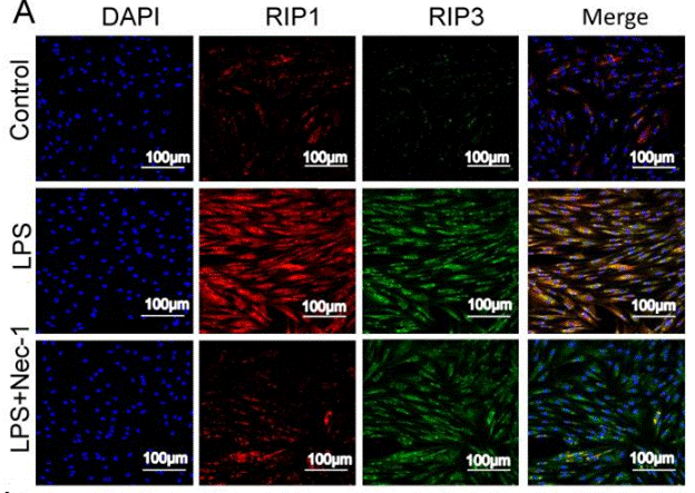
|
30462730 | |
化学信息&溶解度
| 分子量 | 259.33 | 分子式 | C13H13N3OS |
| CAS号 | 4311-88-0 | SDF | Download Necrostatin-1 SDF |
| Smiles | CN1C(=O)C(NC1=S)CC2=CNC3=CC=CC=C32 | ||
| 储存条件(自收到货起) | |||
|
体外溶解度 |
DMSO : 57 mg/mL ( (219.79 mM) ;DMSO吸湿会降低化合物溶解度,请使用新开封DMSO) Water : Insoluble Ethanol : Insoluble |
摩尔浓度计算器 |
|
体内溶解配方 现配现用,请按从左到右的顺序依次添加,澄清后再加入下一溶剂 |
动物体内配方计算器 | |||||
实验计算
动物体内配方计算器(澄清溶液)
第一步:请输入基本实验信息(考虑到实验过程中的损耗,建议多配一只动物的药量)
mg/kg
g
μL
只
第二步:请输入动物体内配方组成(配方适用于不溶于水的药物;不同批次药物配方比例不同,请联系Selleck为您提供正确的澄清溶液配方)
% DMSO
%
% Tween 80
% ddH2O
%DMSO
%
计算结果:
工作液浓度: mg/ml;
DMSO母液配制方法: mg 药物溶于μL DMSO溶液(母液浓度mg/mL,注:如该浓度超过该批次药物DMSO溶解度,请先联系Selleck);
体内配方配制方法:取μL DMSO母液,加入μL PEG300,混匀澄清后加入μL Tween 80,混匀澄清后加入μL ddH2O,混匀澄清。
体内配方配制方法:取μL DMSO母液,加入μL Corn oil,混匀澄清。
注意:1. 首先保证母液是澄清的;
2.一定要按照顺序依次将溶剂加入,进行下一步操作之前必须保证上一步操作得到的是澄清的溶液,可采用涡旋、超声或水浴加热等物理方法助溶。
技术支持
在订购、运输、储存和使用我们的产品的任何阶段,您遇到的任何问题,均可以通过拨打我们的热线电话400-668-6834,或者技术支持邮箱tech@selleck.cn,直接联系到我们。我们会在24小时内尽快联系您。
如果有其他问题,请给我们留言。
* 必填项
Tags: buy Necrostatin-1 | Necrostatin-1 supplier | purchase Necrostatin-1 | Necrostatin-1 cost | Necrostatin-1 manufacturer | order Necrostatin-1 | Necrostatin-1 distributor
