(-)-Blebbistatin
别名: (S)-(-)-Blebbistatin 中文名称:布雷他汀
此产品请避光密封保存。
(-)-Blebbistatin ((S)-(-)-Blebbistatin)是一种细胞渗透性抑制剂,作用于非肌肉肌球蛋白IIATPase,无细胞试验中IC50为~2 μM,不抑制肌球蛋白轻链激酶 (MLCK),抑制卵裂沟的缢缩,不干扰有丝分裂或收缩环的组装。
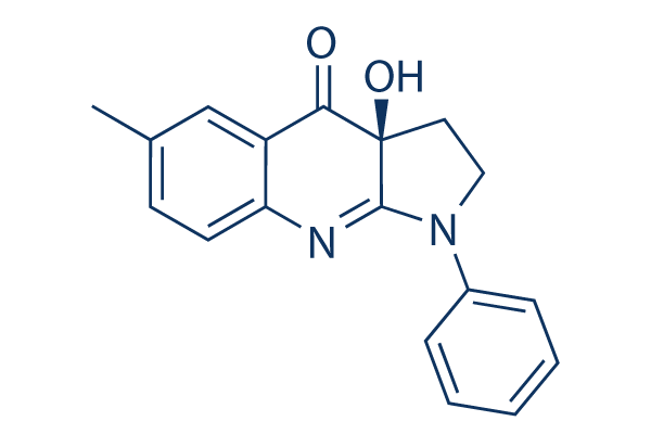
(-)-Blebbistatin Chemical Structure
CAS: 856925-71-8


产品质控
批次:
纯度:
99.82%
99.82
(-)-Blebbistatin相关产品
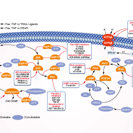
相关信号通路图
细胞实验数据示例
| 细胞系 | 实验类型 | 给药浓度 | 孵育时间 | 活性描述 | 文献信息(PMID) |
|---|---|---|---|---|---|
| Rh41 | qHTS assay | qHTS of pediatric cancer cell lines to identify multiple opportunities for drug repurposing: Confirmatory screen for Rh41 cells | 29435139 | ||
| SK-N-MC | qHTS assay | qHTS of pediatric cancer cell lines to identify multiple opportunities for drug repurposing: Primary screen for SK-N-MC cells | 29435139 | ||
| NB-EBc1 | qHTS assay | qHTS of pediatric cancer cell lines to identify multiple opportunities for drug repurposing: Primary screen for NB-EBc1 cells | 29435139 | ||
| 点击查看更多细胞系数据 | |||||
生物活性
| 产品描述 | (-)-Blebbistatin ((S)-(-)-Blebbistatin)是一种细胞渗透性抑制剂,作用于非肌肉肌球蛋白IIATPase,无细胞试验中IC50为~2 μM,不抑制肌球蛋白轻链激酶 (MLCK),抑制卵裂沟的缢缩,不干扰有丝分裂或收缩环的组装。 | ||
|---|---|---|---|
| 靶点 |
|
| 体外研究(In Vitro) | ||||
| 体外研究活性 | Blebbistatin抑制细胞分裂,改变鱼角膜细胞的平滑运动,且抑制缺乏细丝蛋白的细胞系进行自发起泡。Blebbistatin抑制非肌肉肌球蛋白IIA,非肌肉肌球蛋白IIB,和兔骨骼肌肌球蛋白S1的HMM片段的酶活性,而不抑制平滑肌球蛋白。 Blebbistatin快速且可逆抑制Mg-ATPase活性,也抑制非肌肉肌球蛋白IIA和IIB的体外活性,而极其微弱抑制平滑肌肌球蛋白(IC50=80 μM)。Blebbistatin有效抑制Dictyostelium肌球蛋白II,但极其微弱抑制Acanthamoeba肌球蛋白II。Blebbistatin不抑制代表性的I,V,和X型肌球蛋白超家族成员。 Blebbistatin不与核苷酸竞争性结合到骨骼肌肌球蛋白亚片段-1。Blebbistatin优先结合到ATP酶,与ADP和磷酸盐在活性位点相互作用,减慢磷释放。Blebbistatin既不干扰肌球蛋白与肌动蛋白结合,也不干扰ATP诱导的肌动球蛋白解离。 |
|||
|---|---|---|---|---|
| 实验图片 | 检测方法 | 检测指标 | 实验图片 | PMID |
| Western blot | talin 1 / vinculin / paxillin paxillin / pY31 paxillin / pY397FAK / FAK PY epitopes / vinculin / paxillin |

|
20308429 | |
| Growth inhibition assay | Cell viability Cell death |
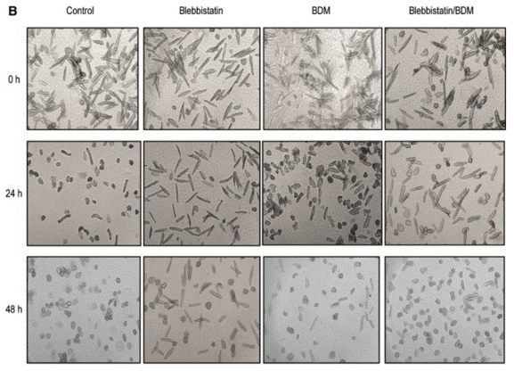
|
26733241 | |
| Glycerol/urea gel electrophoresis | RLC phosphorylation RLC phosphorylation RLC phosphorylation |
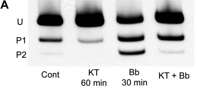
|
18701651 | |
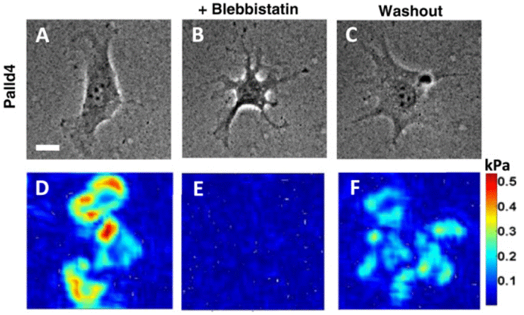
| DIC image | Traction force of a palladin KD (Palld4) cell |
|
27353427 | |
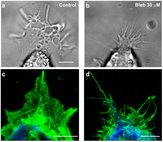
| Immunofluorescence | GCs morphology actin / NMIIA / tubulin VE-cadherin / F-actin PY epitopes / actin PY epitopes / paxillin talin 1 / FAK / β1 integrin / zyxin / vinculin / α-actinin / paxillin |
|
25598228 | |
|
化学信息&溶解度
| 分子量 | 292.33 | 分子式 | C18H16N2O2 |
| CAS号 | 856925-71-8 | SDF | Download (-)-Blebbistatin SDF |
| Smiles | CC1=CC2=C(C=C1)N=C3C(C2=O)(CCN3C4=CC=CC=C4)O | ||
| 储存条件(自收到货起) | 3年 -20°C(避光) 粉状 | ||
|
体外溶解度 |
DMSO : 39 mg/mL ( (133.41 mM) ;DMSO吸湿会降低化合物溶解度,请使用新开封DMSO) Water : Insoluble Ethanol : Insoluble |
摩尔浓度计算器 |
|
体内溶解配方 现配现用,请按从左到右的顺序依次添加,澄清后再加入下一溶剂 |
动物体内配方计算器 | |||||
实验计算
动物体内配方计算器(澄清溶液)
第一步:请输入基本实验信息(考虑到实验过程中的损耗,建议多配一只动物的药量)
mg/kg
g
μL
只
第二步:请输入动物体内配方组成(配方适用于不溶于水的药物;不同批次药物配方比例不同,请联系Selleck为您提供正确的澄清溶液配方)
% DMSO
%
% Tween 80
% ddH2O
%DMSO
%
计算结果:
工作液浓度: mg/ml;
DMSO母液配制方法: mg 药物溶于μL DMSO溶液(母液浓度mg/mL,注:如该浓度超过该批次药物DMSO溶解度,请先联系Selleck);
体内配方配制方法:取μL DMSO母液,加入μL PEG300,混匀澄清后加入μL Tween 80,混匀澄清后加入μL ddH2O,混匀澄清。
体内配方配制方法:取μL DMSO母液,加入μL Corn oil,混匀澄清。
注意:1. 首先保证母液是澄清的;
2.一定要按照顺序依次将溶剂加入,进行下一步操作之前必须保证上一步操作得到的是澄清的溶液,可采用涡旋、超声或水浴加热等物理方法助溶。
技术支持
在订购、运输、储存和使用我们的产品的任何阶段,您遇到的任何问题,均可以通过拨打我们的热线电话400-668-6834,或者技术支持邮箱tech@selleck.cn,直接联系到我们。我们会在24小时内尽快联系您。
如果有其他问题,请给我们留言。
* 必填项


